Meng, Xiangxi
孟祥溪
Research
I am an interdisciplinary researcher working at the intersection of nuclear medicine, molecular imaging, and biomedical engineering. My research focuses on advancing PET imaging technology, developing novel molecular probes, and applying data science to clinical imaging problems. I am particularly interested in the engineering aspects of molecular imaging—including imaging system construction, radiotracer development, and quantitative imaging analysis.
I collaborate closely with physicians, chemists, data scientists, and engineers to translate imaging innovations from bench to bedside, with the ultimate goal of improving patient care through better imaging tools and techniques.
Navigation
Total-body PET imaging for pharmaceutical research
Total-body PET scanners represent a paradigm shift in nuclear medicine imaging. With dramatically increased sensitivity and axial field-of-view, these systems enable new applications in drug development and evaluation that were previously impractical.
My research in this area focuses on:
- Leveraging total-body PET for quantitative pharmacokinetic studies in drug development
- Calibrating the accuracy and precision in quantitative measurement of PET
- Developing novel imaging protocols for multi-organ drug distribution studies
- Application of total-body PET in early phase clinical trials
Key Publications
- Meng, X., Kong, X., Wu, R., & Yang, Z. (2025). Total body PET/CT: A role in drug development? Seminars in Nuclear Medicine, 55(1), 116-123.
- Meng, X., Kong, X., Xia, L., Wu, R., Zhu, H., & Yang, Z. (2024). The role of total-body PET in drug development and evaluation: status and outlook. Journal of Nuclear Medicine, 65(Supplement 1), 46S-53S.
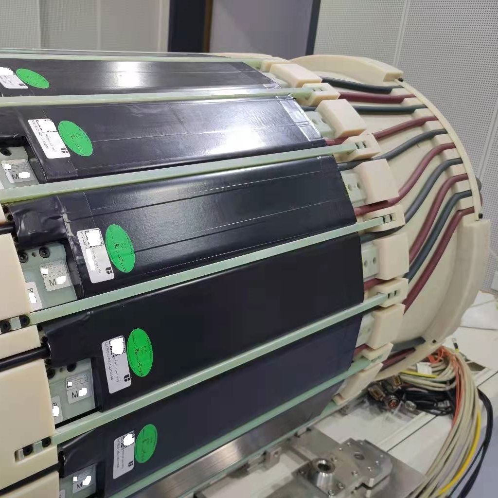
PET imaging physics and instrumentation
The quantitative accuracy and diagnostic utility of PET imaging are fundamentally governed by the underlying physics principles and instrumentation design. My research focuses on comprehensive characterization and optimization of PET system performance to establish rigorous standards for clinical and research applications.
My work in this area encompasses:
- Multi-center harmonization of standardized uptake values (SUVs)
- Systematic performance characterization of integrated PET/MR systems
- Investigation of magnetic field interactions with positron annihilation physics and spatial resolution degradation
- Instrumentation optimization for enhanced quantitative precision in hybrid imaging modalities
Key Publications
- Meng, X., Liu, H., Li, H., Wang, S., Sun, H., Wang, F., Ding, J., He, L., Chen, X., Jin, L., Dong, Y., Zhu, H., & Yang, Z. (2022). Evaluating the impact of different positron emitters on the performance of a clinical PET/MR system. Medical Physics, 49(4), 2642-2651.
- Song, Y., Meng, X., Cao, Z., Zhao, W., Zhang, Y., Guo, R., Zhou, X., Yang, Z., & Li, N. (2023). Harmonization of standard uptake values across different positron emission tomography/computed tomography systems and different reconstruction algorithms: validation in oncology patients. EJNMMI Physics, 10, 19.
Dynamic PET and parametric imaging
Dynamic PET imaging involves continuous acquisition after injection of radiotracers, providing valuable quantitative data for kinetic modeling. This technique reveals pharmacokinetic characteristics and in vivo receptor binding behavior through analysis of 4D data.
My research in this area includes:
- Study of tracer kinetic characteristics and receptor binding with dynamic PET
- Development of novel analysis pipelines for 4D PET data
- Parametric imaging based on multi-modal information fusion
- Patlak graphical plot analysis for reversible tracers
Key Publications
- Shao, W., Zhang, Y., Du, F., Cheng, F., Chen, Y., Meng, X., Liang, Y., & Xie, Z. (2025). Neural network-aided unsupervised input function estimation for dual-time-window PET Patlak analysis. EJNMMI Physics, 12, 100.
- Mao, X., Zhao, S., Meng, X., Kong, H., Yuan, J., He, Q., Liang, D., Yu, J., & Hu, Z. (2022). PET parametric imaging based on MR frequency-domain texture information. Nuclear Instruments and Methods in Physics Research, A, 1029, 166411.

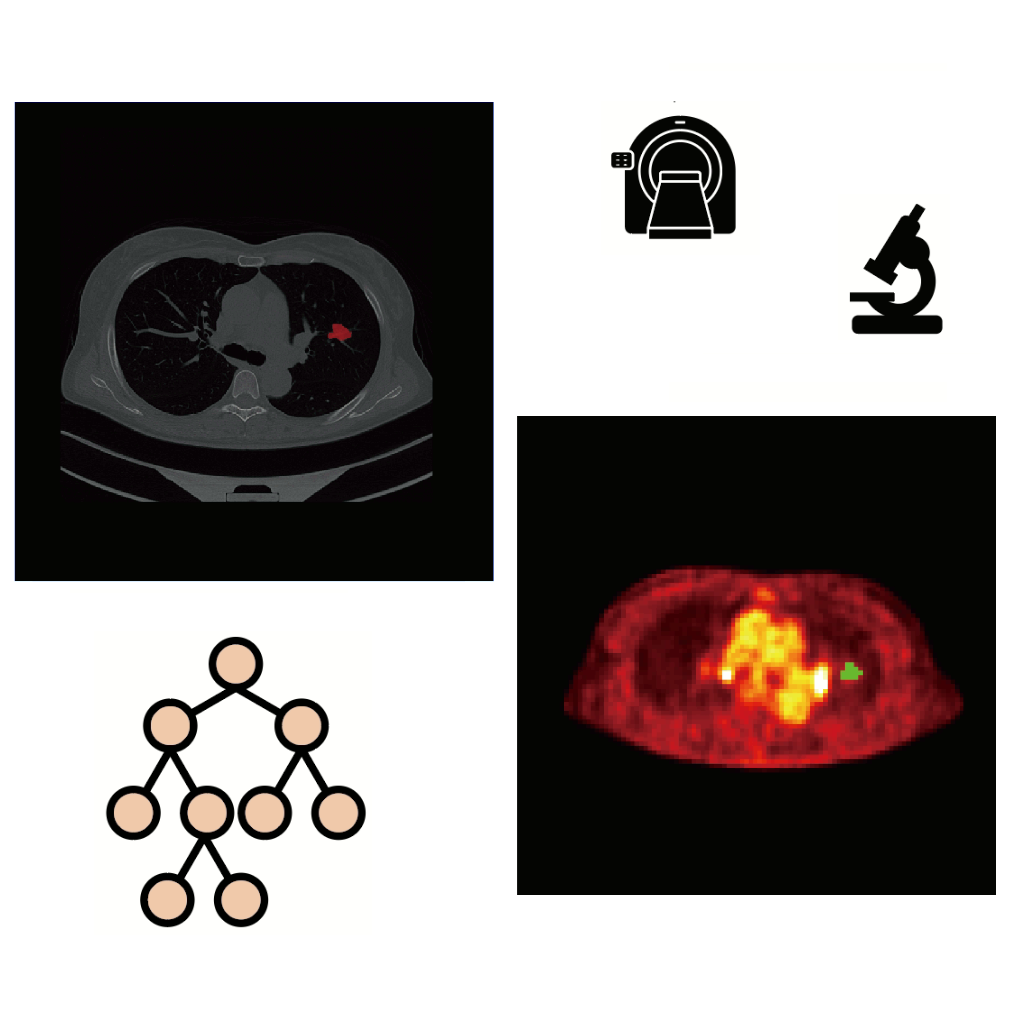
Automation of PET diagnostic pipeline
Machine learning and artificial intelligence are transforming medical imaging by enabling automated analysis and decision support. I work on developing automated diagnostic pipelines for clinical PET applications, with particular focus on lung cancer diagnostics.
My research in this area focuses on:
- Development of automated feature extraction and diagnostic pipelines for PET/CT
- Machine learning-based prediction of pleural invasion in lung cancer using 18F-FDG PET/CT
- Clinical validation and translation of AI-assisted diagnostic tools
- Integration of radiomics with clinical decision-making workflows
Key Publications
- Kong, X., Zhang, A., Zhou, X., Zhao, M., Liu, J., Zhang, X., Zhang, W., Meng, X., Li, N., & Yang, Z. (2025). A strategy for the automatic diagnostic pipeline towards feature-based models: a primer with pleural invasion prediction from preoperative PET/CT images. EJNMMI Research, 15, 70.
- Zhang, A., Zhao, M., Kong, X., Zhang, W., Hou, X., Yang, Z., Meng, X., & Li, N. (2025). The predictive value of 18F-FDG PET/CT radiomics for pleural invasion in non-small cell lung cancer. European Journal of Radiology, 190, 112199.
- Zhang, A., Meng, X., Yao, Y., Zhou, X., Yan, S., Wang, F., Zhou, N., Zhang, Y., Kong, H., & Li, N. (2023). Predictive value of 18F-FDG PET/MRI for pleural invasion in solid and subsolid lung adenocarcinomas smaller than 3 cm. Journal of Magnetic Resonance Imaging, 57(5), 1367-1375.
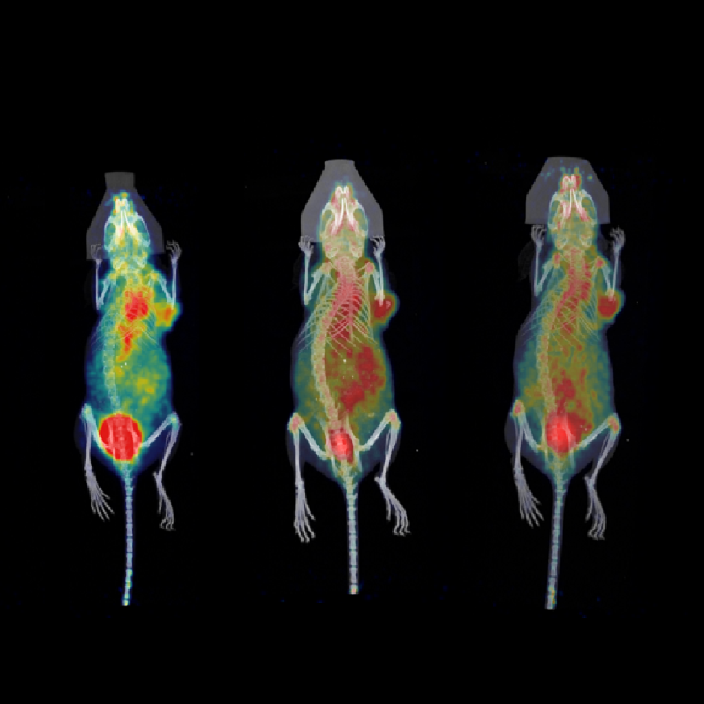
Molecular probes and tracers
Molecular imaging probes serve as the bridge between biological targets and imaging signals. I have been involved in developing novel radiotracers and molecular probes for imaging specific molecular targets, with particular focus on nanobody-based agents.
My tracer development research focuses on:
- Radiopharmaceutical development based on nanobodies
- Developing tracers with potential diagnostic and therapeutic applications
- Preclinical evaluation and pilot clinical translation studies
- Application of pharmacological methods in drug development
Key Publications
- Zheng, D., Huang, Y., Li, C., Lu, Y., Xie, Z., Ren, Q., Liang, Y., & Meng, X. (2025). A Radiofluorinated nanobody PET tracer to dynamically visualize differential Nectin4 expression in vivo. Journal of Medicinal Chemistry, 68(20), 21366-21376.
- Zheng, D., Huang, Y., Li, C., Lu, Y., Xie, Z., Liang, Y., Ren, Q., & Meng, X. (2025). Novel radiofluorinated nanobody PET tracer for preclinical studies of TIM3 expression. Molecular Pharmaceutics, 22(10), 6141-6150.